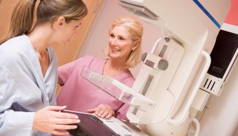
Let’s talk about mammograms. Mammograms are an essential tool for the early detection and prevention of breast cancer. It is the best way to discover breast cancer early and is the only way to detect lumps or masses that are too small to be felt. Right now, mammograms are the most effective tool at our disposal for detecting and treating breast cancers early. It is recommended by most medical organizations that women with an average risk of breast cancer consider regular mammogram testing starting at age 40 and repeating the screening annually. Digital mammograms – the most common type – are an effective screening tool for women with or without dense breast tissue since they allow for a more detailed analysis. You and your physician may consider supplemental testing based on other risk factors and personal preferences. Here are a few things you need to know about mammograms.
Who Should Get a Mammogram?
The American Cancer Society recommends the following guidelines for women at average risk:
- Women between 40 and 44 can start screening with a mammogram every year.
- Women 45 to 54 should get mammograms every year.
- Women 55 and older can switch to a mammogram every other year or choose to continue yearly mammograms. Screening should continue if a woman is in good health and is expected to live at least ten more years.
The American Cancer Society also recommends that women at high risk should get a mammogram yearly. Women who are at increased risk for breast cancer can include any of the following:
- Those with a family history of breast cancer
- Those with a personal history of breast cancer, ductal carcinoma in situ (DCIS), lobular carcinoma in situ (LCIS), atypical ductal hyperplasia (ADH), or atypical lobular hyperplasia (ALH)
- Those who had radiation therapy on their chest between 10 and 30 years of age
- Those with a known BRCA1 or BRCA2 gene mutation
- Those who have not had genetic testing themselves but have a first-degree relative (parent, sibling, or child) with a known BRCA1 or BRCA2 gene mutation
- Those with Li-Fraumeni syndrome, Cowden syndrome, or Bannayan-Riley-Ruvalcaba syndrome have first-degree relatives with one of these syndromes.
What is a Mammogram?
There are several different mammograms, but a mammogram is essentially an x-ray of the breast to detect tumors and abnormalities that could be cancerous. Almost all mammography done in the United States is digital instead of conventional or film mammography. With conventional mammography, the breast image was saved on film. With digital mammography, the digital image is saved as a computer file, allowing doctors to share the file or access it remotely, making it faster and easier to diagnose the image. In addition to regular digital mammography, which produces a two-dimensional breast image, 3-D mammography or tomosynthesis is now available. 3-D mammography uses low-dose X-rays to take multiple breast images at different angles and uses special software to put them together into a 3-D image. The 3-D images are superior to those from a traditional digital image which, is hoped anyway, will help catch more cancers and reduce unnecessary biopsies and follow-ups.
Moreover, a recent study suggests that about half of women who routinely get mammograms haven’t heard of the term “baseline mammogram,” a recent study suggests. So, what is a baseline mammogram? Simply put, it’s a person’s first mammogram. There are several things that a breast radiologist looks for when interpreting a mammogram, including an interval change in the appearance of the breasts since prior mammograms. So, once a baseline mammogram is taken, it serves as a comparison for any subsequent exam.
Do you have dense breast tissue? Would you know what it meant if you did?
For those who aren’t sure what dense breast tissue is, it refers to the appearance of breast tissue on a mammogram; it’s normal and a common finding. Knowing whether you do or do not have dense breasts is a critical factor in the early detection of breast cancer. The density of your breasts helps determine whether a mammogram will be enough to detect breast cancer or if you may need supplementary screenings. Once your mammogram is completed, the radiologist who conducted the mammogram will determine the ratio of non-dense tissue and dense tissue and assign a breast density level. The radiologist typically will use a results reporting system called Breast Imaging Reporting and Data System (BI-RADS). The levels of density frequently are recorded using a letter:
A.) Almost entirely fatty. This indicates the breasts are almost entirely made of fat. About 1 in 10 women have this result.
B.) Scattered area of fibroglandular density. This means there are some scattered areas of density, but most of the tissue is nondense. This happens to about 4 in 10 women.
C.) Heterogeneously dense. This indicates that there are areas of nondense tissue, but most of the breast tissue is dense. About 4 in 10 women have this result.
D.) Extremely dense. This indicates that nearly all of the tissue is dense. Only about 1 in 10 women have this result.
Women are considered to have dense breasts when they are classified as heterogeneously dense or extremely dense. Almost half of women aged 40 and older who undergo mammograms are found to have dense breasts. They are often inherited, but some factors can influence dense breasts. If you have a lower body mass index, you are more likely to have dense breast tissue than women who are obese. You may also have a higher chance of dense breast tissue if you take hormone therapy for menopause. If you have dense breast tissue, your doctor may recommend other supplemental tests for breast cancer screenings, including:
- 3-D Mammogram or Breast Tomosynthesis. This screening uses X-rays to collect several images of the best from multiple angles. A computer then synthesizes the images to create a 3-D image of the breast.
- Breast MRI. MRIs use magnets to create breast images and don’t use radiation. This screening is recommended for women with a very high risk of breast cancer.
- Breast Ultrasound. This screening uses sound waves to analyze breast tissue and is commonly used to investigate areas of concern discovered on a mammogram.
- Molecular Breast Imaging or BMI. Also known as breast-specific gamma imaging, uses a special gamma camera that records the activity of a radioactive tracer. This tracer is injected into a vein in the arm where it can see the difference between normal tissue and cancerous tissue because they react differently to the reactor. These reactions can be seen in the images created by the gamma camera. MBI is performed every other year in addition to an annual mammogram.
When to Get a Mammogram?
The risk for breast cancer increases with age. The recommendation is for all women to begin yearly mammographic screening at age 40. For women with a strong family history of breast cancer, a history of radiation therapy to the chest, or a known genetic mutation, screening may begin at an earlier age, and they should consult their physician.
If you are at higher risk due to genetic or other factors, please consult with your doctor as to when you should start screening (which is often before age 40). Talk with your doctor about your personal history, family health history, and your risk factors and together as a team decide what’s best for you.
The American Cancer Society also recommends scheduling your mammogram for about a week after your menstrual period, if possible. Mammograms can be uncomfortable and sometimes painful. Your breasts will be less swollen and tender a week after your menstrual period, which means less pain and discomfort during the mammogram.
Tips for Getting a Mammogram
• Try to avoid having your mammogram the week before you get your period or during your period, if possible. Mammograms can be uncomfortable and sometimes painful. Your breasts will be less swollen and tender a week after your menstrual period, which means less pain and discomfort during the mammogram.
• On the day of your mammogram, don’t wear deodorant, perfume, or powder. These products can show up as white spots on the X-ray.
• Some women prefer to wear a top with a skirt or pants instead of a dress. You will need to undress from your waist up for the mammogram.
Where to get a Mammogram?
Community Care Physicians is proud to have The Breast Center at ImageCare, one of the Capital Region’s few full-service breast centers providing the latest breast cancer screenings and diagnosis. Our accredited facility offers state-of-the-art technology paired with excellent patient care to serve women’s breast health needs. Our subspecialized radiologists and team of specialists are board-certified in breast imaging and leaders in their specialized fields in administering and reading mammography, breast ultrasound, and breast MRI.
The Breast Center at ImageCare not only specializes in and performs breast screenings but also provides patients access to our unique Oncology Nurse Navigator to help patients navigate the healthcare landscape for cancer screenings, diagnosis, treatment, and support services. The Breast Center has two full-service locations in Latham and Clifton Park, and ImageCare Medical Imaging also provides mammography services in Saratoga, Balltown, and Guilderland. Click here to view The Breast Center at ImageCare’s website and learn more about their services and specialized and dedicated staff.
Why Get a Mammogram?
Mammograms save lives. Mammography is the most effective tool available today for screening to find breast cancer. Mammography correctly detects 87% of women with breast cancer that would otherwise go undiagnosed. Breast Cancer is the most commonly diagnosed cancer in the United States, combined with the increased risk after age 45. The overall effectiveness of mammograms is why every woman over 45 should get a yearly mammogram and why every woman over 40 should consider getting a screening mammogram every year.
Not all mammograms are routine screenings. If you or your doctor feels a lump or notices any changes in the appearance or feel of your breasts, talk to your doctor about getting a diagnostic mammogram. Symptoms such as changes in breast size and shape, a lump in the breast, swelling in the armpit, pain or swelling of the breast, nipple changes or discharge, and certain high-risk factors may be a reason for your doctor to recommend a diagnostic mammogram.
